FIGURE

Figure 3

ID
ZDB-FIG-220801-55
Publication
Lin et al., 2022 - Quantification of Idua Enzymatic Activity Combined with Observation of Phenotypic Change in Zebrafish Embryos Provide a Preliminary Assessment of Mutated idua Correlated with Mucopolysaccharidosis Type I
Other Figures
All Figure Page
Back to All Figure Page
Figure 3

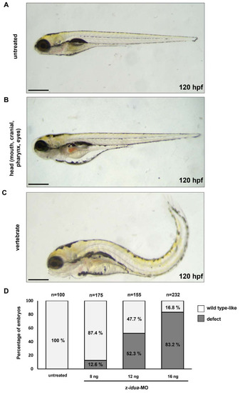
Knockdown of z-idua caused defective development in zebrafish embryos. Embryonic phenotypes were observed in 120 hpf zebrafish embryos injected with 12 ng z-idua-MO. (A) Untreated control embryos; (B,C): z-idua-MO-injected embryos; (B) defective head, including smaller eyes, abnormal craniofacial and pharyngeal arches, and cardiac edema; (C) defective body shape, including shortened somites and bent body axis. (D) Percentage of defective phenotypes occurring in all examined embryos injected with z-idua-MO. Embryonic morphology was examined at 120 hpf in the zebrafish embryos injected at one-cell stage with different concentrations of z-idua-MO, as indicated. The occurrence rates (in percentages) of defective phenotypes (marked in black boxes) and the wild type-like phenotype (marked in grey) among the total examined embryos injected were calculated. The total number (n) of embryos studied in each group was indicated at the top of each column. Scale bar: 500 μm.

Expression Data

Expression Detail
Antibody Labeling
Phenotype Data

Phenotype Detail
Acknowledgments
This image is the copyrighted work of the attributed author or publisher, and ZFIN has permission only to display this image to its users. Additional permissions should be obtained from the applicable author or publisher of the image. Full text @ J Pers Med