FIGURE

Figure 3

ID
ZDB-FIG-220717-60
Publication
Lucas et al., 2022 - Pannexin 1 drives efficient epithelial repair after tissue injury
Other Figures
All Figure Page
Back to All Figure Page
Figure 3

Macrophages expand and augment epithelial proliferation in response to tissue injury.

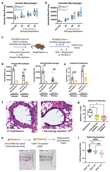
(a) Lung alveolar macrophage numbers (CD45+/CD11c+/Siglec-F+ cells) and (b) interstitial macrophage numbers (CD45+/CD11b+/CD64+ cells) in digested right lung lobes after naphthalene-induced epithelial injury (200mg/kg) analysed by flow cytometry of lung digests in Panx1+/+ and Panx1-/- mice (n=3-5 corn oil; n=5-9 post-naphthalene). (c) Schema of experimental protocol for lung macrophage depletion in Panx1+/+ mice, achieved by intratracheal (i.t.) administration of 100μL clodronate liposomes to deplete alveolar macrophages, and PLX5622 containing chow (1200mg/kg feed) ad libitum for 3d prior to experimentation with additional PLX5622 (65mg/kg) given by oral gavage (on days 0,1,2,3&4 of naphthalene administration) to deplete interstitial macrophages. (d) Confirmation of macrophage depletion after epithelial injury and (e) In vivo epithelial injury-induced proliferation analysed by EdU incorporation (EdU 1.5mg/mouse given intraperitoneally (i.p.) on days 4&4.5) with or without macrophage depletion in response to high dose (200mg/kg, n=3) or intermediate dose (160mg/kg n=8) naphthalene, assessed by t-test. (f) Representative H&E staining of lung tissue sections at day 7 after naphthalene injury (200mg/kg) with or without macrophage depletion, with injured and repairing airway epithelium highlighted by blue arrows. Grey scale bars, 50um. (g) In vivo epithelial injury-induced proliferation analysed by EdU incorporation with alveolar macrophage depletion (by clodronate liposomes) or interstitial macrophage depletion (by PLX5622) in response to naphthalene (160mg/kg, n=4/group, one experiment, assessed by one-way ANOVA with Holm-Šídák's multiple comparisons test). (h) Macrophage ablation achieved using the csf1ra:NfsB line (NfsB) and metronidazole (met) treatment (NfsB+/met-, NfsB-/met+ and NfsB+/met+ groups; macrophage depletion only with NfsB+/met+) with macrophage ablation confirmed in NfsB+/met+ fish by fluorescent microscopy (mCherry+ macrophages pseudocoloured in magenta) scale bar 100um. (i) Defective tailfin regeneration in macrophage-deficient zebrafish (NfsB+/met+ group) (n=14, assessed by one-way ANOVA with Holm-Šídák's multiple comparisons test). *p<0.05, **p<0.01 *** p<0.001, ****p<0.0001.

Expression Data

Expression Detail
Antibody Labeling
Phenotype Data

Phenotype Detail
Acknowledgments
This image is the copyrighted work of the attributed author or publisher, and ZFIN has permission only to display this image to its users. Additional permissions should be obtained from the applicable author or publisher of the image. Full text @ Sci Immunol