FIGURE SUMMARY
Title

Pannexin 1 drives efficient epithelial repair after tissue injury

Authors
Lucas, C.D., Medina, C.B., Bruton, F.A., Dorward, D.A., Raymond, M.H., Tufan, T., Etchegaray, J.I., Barron, B., Oremek, M.E.M., Arandjelovic, S., Farber, E., Onngut-Gumuscu, S., Ke, E., Whyte, M.K.B., Rossi, A.G., Ravichandran, K.S.
Source
Full text @ Sci Immunol

Pannexin1 is required for efficient epithelial repair after tissue injury.

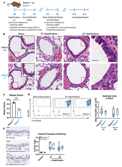
(a) Wild type (Panx1+/+) or pannexin 1 globally deficient mice (Panx1-/-) were injected with naphthalene (200mg/kg) i.p. to cause acute airway epithelial death prior to analysis of bronchoalveolar lavage fluid (BALF) or lung tissue. (b) Representative H&E staining of lung tissue sections from naïve Panx1+/+ or Panx1-/- mice and at day 1 (D1) and D7 post-naphthalene injury, with injured and repairing airway epithelium highlighted by yellow arrows. Grey scale bars, 50μm; black scale bars, 20μm. Dotted red lines reflect insets in naïve images and zoomed in images in D7 naphthalene images. (c) H&E sections were blinded prior to analysis by a lung pathologist (n=3 per group, assessed by t-test, one independent experiment). (d) BALF was acquired at day 1 or day 2 post-naphthalene injury and cellular content analysed for epithelial cells (CD45-/EpCAM+ cells) and their staining with a live-dead (LD) marker (low staining of LD marker are live cells, high staining are dead cells; n=4-6, two independent experiments). (e) Caspase-3 activation was assessed by cleaved caspase-3 staining on lung tissue sections in the absence of injury and on days 1 and 2 after naphthalene epithelial injury (n=4-7, two independent experiments). Black scale bars, 20um. **p<0.01.

Pannexin1 promotes injury-induced epithelial proliferation, and boosts tailfin regeneration in zebrafish.

(a-c) In vivo epithelial injury-induced proliferation in response to naphthalene was analysed by EdU incorporation of CD45-/CD31-/EpCAM+ cells (n=3-8, analysed by one-way ANOVA with Dunnett's multiple comparisons test). (d,e) Injury-induced epithelial proliferation was measured in Panx1+/+ and Panx1-/- mice, with naïve mouse data shown for comparison (n=3-6 naïve, n=5-8 post-naphthalene, two independent experiments, assessed by t-test). (f) Zebrafish at 3 days post-fertilisation were microinjected with 1nL of the Panx1 pharmacological inhibitors trovafloxacin (80mM) or spironolactone (120mM), or DMSO control, and tailfins were transected. (g,h) Tailfin regeneration rate was assessed at 48h (n=20-22 separate animals, three separate experiments, assessed by one-way ANOVA with Holm-Šídák's multiple comparisons test). Scale bar, 100um. (i) Proliferation in the regenerating tailfin analysed by EdU incorporation, imaged by light sheet fluorescent microscopy (n=15-21 per group, assessed by Holm-Šídák's multiple comparisons test). (j) Similarly, Panx1a morphants (or control sequence morphants) underwent tailfin transection and regeneration rate was assessed at 48h post-injury (n=13 separate animals, two separate experiments, assessed by t-test), with (k) tailfin proliferation analysed by EdU incorporation, (n=21 per group, one experiment, assessed by t-test). *p<0.05, ***p<0.001, **** p<0.0001.

Macrophages expand and augment epithelial proliferation in response to tissue injury.

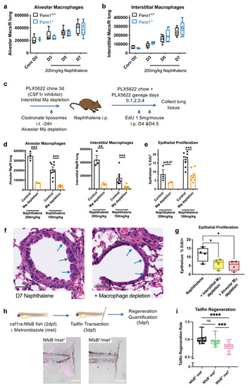
(a) Lung alveolar macrophage numbers (CD45+/CD11c+/Siglec-F+ cells) and (b) interstitial macrophage numbers (CD45+/CD11b+/CD64+ cells) in digested right lung lobes after naphthalene-induced epithelial injury (200mg/kg) analysed by flow cytometry of lung digests in Panx1+/+ and Panx1-/- mice (n=3-5 corn oil; n=5-9 post-naphthalene). (c) Schema of experimental protocol for lung macrophage depletion in Panx1+/+ mice, achieved by intratracheal (i.t.) administration of 100μL clodronate liposomes to deplete alveolar macrophages, and PLX5622 containing chow (1200mg/kg feed) ad libitum for 3d prior to experimentation with additional PLX5622 (65mg/kg) given by oral gavage (on days 0,1,2,3&4 of naphthalene administration) to deplete interstitial macrophages. (d) Confirmation of macrophage depletion after epithelial injury and (e) In vivo epithelial injury-induced proliferation analysed by EdU incorporation (EdU 1.5mg/mouse given intraperitoneally (i.p.) on days 4&4.5) with or without macrophage depletion in response to high dose (200mg/kg, n=3) or intermediate dose (160mg/kg n=8) naphthalene, assessed by t-test. (f) Representative H&E staining of lung tissue sections at day 7 after naphthalene injury (200mg/kg) with or without macrophage depletion, with injured and repairing airway epithelium highlighted by blue arrows. Grey scale bars, 50um. (g) In vivo epithelial injury-induced proliferation analysed by EdU incorporation with alveolar macrophage depletion (by clodronate liposomes) or interstitial macrophage depletion (by PLX5622) in response to naphthalene (160mg/kg, n=4/group, one experiment, assessed by one-way ANOVA with Holm-Šídák's multiple comparisons test). (h) Macrophage ablation achieved using the csf1ra:NfsB line (NfsB) and metronidazole (met) treatment (NfsB+/met-, NfsB-/met+ and NfsB+/met+ groups; macrophage depletion only with NfsB+/met+) with macrophage ablation confirmed in NfsB+/met+ fish by fluorescent microscopy (mCherry+ macrophages pseudocoloured in magenta) scale bar 100um. (i) Defective tailfin regeneration in macrophage-deficient zebrafish (NfsB+/met+ group) (n=14, assessed by one-way ANOVA with Holm-Šídák's multiple comparisons test). *p<0.05, **p<0.01 *** p<0.001, ****p<0.0001.

Pannexin1 at the site of tissue injury regulates epithelial injury-induced proliferation.

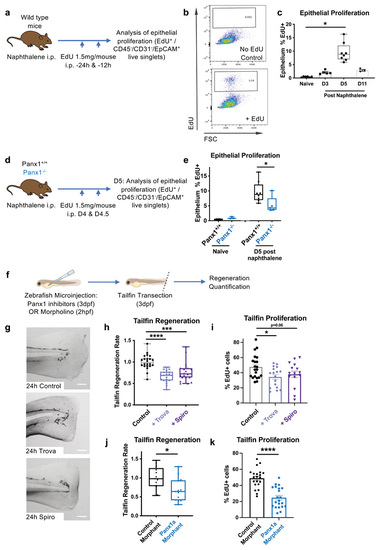
(a) Schema of experimental protocol for deleting Panx1 using CX3Cr1-Cre or LysM-Cre mediated approaches prior to epithelial injury, with (b) in vivo epithelial injury-induced proliferation analysed by EdU incorporation (n=4-5/group Cx3Cr1 Cre, assessed by t-test; n=4/group LysM Cre, assessed by t-test). (c) Schema of local delivery of TAT-Cre into the lungs. (d) Representative flow cytometry plots of YFP expression within epithelium with either PBS (vehicle) or TAT-Cre delivery with (e) confirmation of TAT-Cre-dependent YFP expression within epithelium and (f) analysis of major immune cell populations in response to TAT-Cre. (g) Schema of experimental protocol for expressing Panx1 transgene (Tg) on a global Panx1-deficient mouse (Panx1-/-) using i.t. TAT-Cre administration. (h) Panx1 protein expression on whole lung extracts analysed in Panx1-/-/Tg- and Panx1-/-/Tg+ mice with and without local TAT-Cre, confirming Panx1 expression in a Tg-dependent and Cre-dependent fashion. (i) In vivo epithelial injury-induced proliferation (analysed by EdU incorporation post-naphthalene) is boosted by re-expression of Panx1 at the site of tissue injury (n=3/group, one experiment, assessed by t-test) with both genotypes treated intra-tracheally with TAT-Cre. *p<0.05.

Dying cell pannexin1 imprints an injury phenotype upon macrophages.

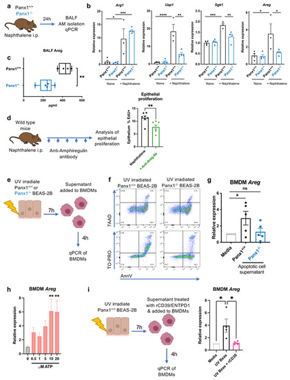
(a) Schema of experimental protocol for analysis of gene expression in lung alveolar macrophages at day 1 after naphthalene-induced epithelial injury in Panx1+/+ and Panx1-/- mice. (b) qPCR analysis of Arg1, Uap1, Sgk1 and Areg in naïve and post-naphthalene injury alveolar macrophages from Panx1+/+ and Panx1-/- mice (n-3-4/group, one experiment, assessed by one-way ANOVA with Tukey's multiple comparisons test). (c) Analysis of amphiregulin protein in BALF supernatant in Panx1+/+ and Panx1-/- mice at day 1 after epithelial injury, analysed by Luminex (n=4-5/group, one experiment, assessed by t-test). (d) Schematic for neutralising amphiregulin in vivo by administration of anti-amphiregulin antibody (5μg in 200μL sterile PBS given intraperitoneally) on days 0,1,2,3,4,&4.5 after naphthalene injury, with epithelial proliferation assessed by EdU incorporation (n=7/group, two independent experiments, assessed by t-test). (e) Schema of experimental protocol for inducing apoptosis in Panx1+/+ or Panx1-/- BEAS-2B epithelial cells with (f) confirmation of apoptosis induction (AnnV/7AAD staining) and Panx1 channel opening in Panx1+/+ apoptotic cells (AnnV/TO-PRO staining) and (g) apoptotic cell supernatant transferred onto mouse bone marrow derived macrophages (BMDMs) for 4h prior to qPCR for Areg (n=5, assessed by oneway ANOVA with Holm-Šídák's multiple comparisons test). (h) BMDMs were treated with increasing concentrations of the Panx1-released nucleotide ATP for 4h prior to qPCR for Areg (n=4, assessed by one-way ANOVA with Holm-Šídák's multiple comparisons test, each group compared to control/0μM ATP). (i) Degradation of extracellular ATP using recombinant CD39/ENTPD1 (rCD39; 44ng/ml) attenuates induction of Areg by BMDMs in response to apoptotic epithelial cell supernatants (qPCR after 4h; n-=4, assessed by one-way ANOVA with Holm-Šídák's multiple comparisons test). *p<0.05, **p<0.01. *** p<0.001, **** p<0.0001.

Pannexin 1 functions upstream of epithelial Nras and Bcas2 to drive injury-induced proliferation in the regenerating epithelium.

(a) Schema of experimental protocol for RNAseq analysis of purified lung epithelium from Panx1+/+ and Panx1-/- mice after naphthalene epithelial injury with (b) heat map of differentially regulated genes, units representing log2 scaled counts. (c) CRISPR–Cas9 genetic deletion of Panx1, Nras and Bcas2 was performed in BEAS-2B epithelial cells (confirmation at transcript and protein level) with (d) proliferation of these cell lines measured by EdU incorporation over 24h, displayed relative to control (n=5, assessed by Kruskal-Wallis test with Dunn's multiple comparisons test). (e) Measurement of Nras (n=7) and Bcas2 (n-5) in BEAS-2B cells after overnight treatment with recombinant human amphiregulin (rAreg) (assessed by Wilcoxon signed-rank test). (f,g) Generation of Nras and Bcas2 zebrafish morphants (or control sequence morphants) for assessment of regeneration rate at 48h post-tailfin transection (n=12-18 separate animals, two separate experiments, assessed by t-test) and (h) tailfin proliferation analysed by EdU incorporation (n=14-39 per group, assessed by t-test). *p<0.05, **p<0.01.

Acknowledgments
This image is the copyrighted work of the attributed author or publisher, and ZFIN has permission only to display this image to its users. Additional permissions should be obtained from the applicable author or publisher of the image. Full text @ Sci Immunol