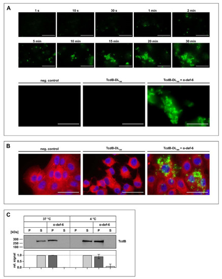
The incubation of α-def-6 and TcdB leads to rapid aggregation. (A) α-def-6 (6 µM) was added to TcdB-DL488 (30 nM) in PBS, and image acquisition with an epifluorescence microscope was started immediately. Aggregation was allowed to proceed for 0.5 h at 37 °C while images were being taken. Representative images of various time points are depicted to show the rapid progression of aggregation over time (upper panel). TcdB-DL488 (30 nM) alone in PBS was used as a positive control, while the same volume of Dylight488 treated PBS in PBS served as a negative control. Images after 0.5 h at 37 °C are depicted (lower panel), (n ≥ 2). (B) Vero cells were intoxicated with TcdB-DL488 (22 nM) in the absence or presence of α-def-6 (6 µM) for 0.5 h at 37 °C. Cells subjected to the same volume of DyLight488-treated PBS, as used TcdB-DL488, served as control. Cells were fixed and permeabilized. Nuclei (blue) were stained with Hoechst33342 and actin (red) with SiR-actin. Representative images taken with an epifluorescence microscope are depicted (n = 2). Scale bars correspond to 50 µm. (C) TcdB (50 ng) was incubated with or without α-def-6 (6 µM) in PBS for 0.5 h at 37 °C. Samples were centrifuged to segregate aggregates and divided into a supernatant (S) and pellet (P) fraction. Fractions were subjected to SDS-PAGE followed by Western blotting. TcdB was detected. Signals were analyzed as the ratio of one fraction (P or S) to the entire sample (P + S) and are depicted as mean ± SD of three biological replicates (n = 3).
|
