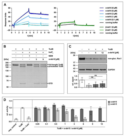
α-def-6 directly binds to TcdB in vitro but does not affect the autoproteolytic- and glucosyltransferase activity of TcdB. (A) 2400 RUs of biotinylated TcdB were immobilized via streptavidin-biotin binding onto a streptavidin sensor chip, and α-def-6 (concentrations as indicated) was applied to the chip for 4 min followed by a dissociation phase with a flow of running buffer. A solution of 6 µM of α-def-6 was injected twice to assess reproducibility, and β-def-1 (6 µM) served as a control. The flow rate was 25 µL/min. (B) The autoproteolytic activity of TcdB (2 µg) in the presence of InsP6 (1 mM) was evaluated in the absence or presence of α-def-6 (3 µM, 9 µM). N-ethylmaleimide (NEM; 1 mM), an inhibitor of the autoproteolytic activity of TcdB, was included as the control. After 1 h incubation at 37 °C, the samples were subjected to SDS-PAGE, and the protein was visualized by Coomassie staining. A representative gel is shown (n = 2). (C) CaCo-2 cell lysate (40 µg) as source of Rac1 was incubated with TcdB (50 ng) in the absence or presence of α-def-6 (6 µM, 12 µM) for 2 h at 37 °C. After SDS-PAGE and Western blotting, non-glucosylated Rac1 was detected with a specific antibody. GAPDH served as the loading control. A representative Western blot is shown. Relative signal intensities normalized to loading control are given as mean ± SD of four biological replicates, each with two technical duplicates (n = 4). Significance was tested with a one-way ANOVA combined with Dunnett’s multiple comparison test (ns = not significant p > 0.05). (D) UDP-GloTM glucosyltransferase assay was performed to analyze the glucosyltransferase activity of TcdB. TcdB (200 pM) was incubated with α-def-5 or α-def-6 (concentrations as indicated) in the presence of recombinant Rac1 (5 µM) as substrate. Castanospermine (Cast; 10 mM), a known inhibitor of the glucosyltransferase activity, was included as a control. Reactions were started with the addition of UDP-glucose (100 µM) and allowed to proceed for 1 h at 37 °C. Samples were combined with UDP detection reagent, and luminescence was measured. Values represent mean ± SD of at least three biological replicates, each with three technical replicates (n ≥ 3).
|
