|
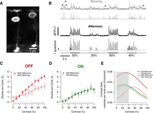
Diurnal modulation of synaptic gain.A Multiphoton section through the eye of a zebrafish larva (7 dpf) expressing iGluSnFR in a subset of bipolar cells. B Examples of iGluSnFR signals from an individual OFF synapse elicited using stimuli of variable contrast modulated at 5 Hz (0–100%, full field, sine wave) in the morning (ZT 0–2 h, grey) and afternoon (ZT 6–8 h, black). Note the high levels of spontaneous activity in the morning (black arrowheads). In each case the top trace shows the iGluSnFR signal and the lower trace the estimated number of quanta composing each event (Qe). C Average contrast-response functions in OFF bipolar cell synapses in the morning (open circles; n = 20 synapses) and afternoon (closed; n = 59), where the response (R) was quantified as the average of quanta per cycle, Qc (i.e., the total number of quanta released within a single cycle of the sinusoidal stimulus). Each point shows the mean ± s.e.m. The smooth lines are fits of a sigmoid used for smoothing. Note the differences in the shape of the contrast-response functions and in the levels of spontaneous activity (zero contrast) (One-way ANCOVA test, p < 0.0006). D Average contrast-response functions in ON bipolar cell synapses in the morning (open circles; n = 12 synapses) and afternoon (closed; n = 31). There was no significant difference in the morning relative to afternoon. (One-way ANCOVA test, p = 0.53) Each point shows the mean ± s.e.m. E The contrast gain calculated as the derivative of the fits to the contrast-response functions in (C, D). The grey box provides an indication of the contrasts most common in nature (below about 40%). Note that the maximum contrast discrimination is increased by a factor of 2x in the OFF channel during the afternoon. Source data are provided as a Source Data file.
|