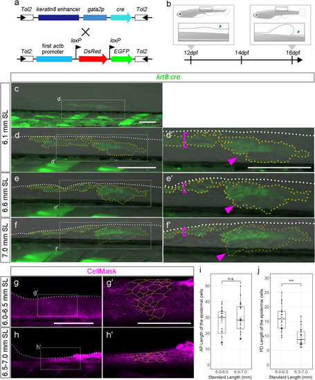
Cell-tracking analysis of the epithelial cells in the reducing LMFF area. (a) Schematic of the plasmid DNA construct used to generate the Tg. (b) Scheme of the Tg observation. (c–f’) GFP-positive labelled cells in the reducing LMFF area at 6.1 mm SL (c–d’), 6.6 mm SL (e–e’), and 7.0 mm SL (f–f’). The right panels (d’,e’,f’) are magnified views of the dashed rectangles in the left panes (d,e,f), respectively. White dashed lines in (d-f’) indicate outlines of the LMFFs. Yellow dashed lines indicate outlines of the EGFP-positive populations of epidermal cells. Magenta brackets in (d’,e’,f’) indicate EGFP-positive populations of epidermal cells experiencing proximo-distal shrinking. Magenta arrowheads in (d’,e’,f’) indicate EGFP-positive populations of epidermal cells migrating down to the trunk. (g–h’) Cell morphology and distribution in the reducing LMFF area at 6.0–6.5 mm SL (g–g’) and 6.5–7.0 mm SL (h–h’). Cell membrane visualized by CellMask. The right panels (g’,h’) are magnified views of the dashed rectangles in the left panels (g,h), respectively. Yellow dashed lines indicate outlines of the epidermal cells. (i,j) Boxplots of cell length along the AP and PD axis in the reducing LMFF area. Whiskers in (i) and (j) show maximum and minimum values within 1.5 times the interquartile range. Boxes show the median and 25th and 75th percentiles. The P value in (i) and (j) is the result of Brunner-Munzel test (P = 0.4407 and P = 8.34e-10). Scale bars in (c,d,d’,g) and that in (g’) indicate 200 μm and 100 μm, respectively.
|
