
Fig. 8
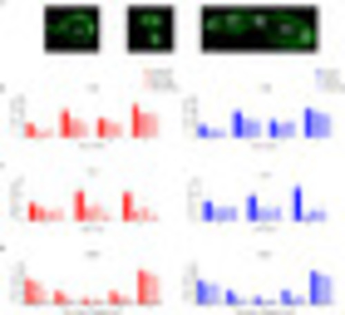
Fig. 8. Number of hair cells in the inner ear organs of adult zebrafish. A) Regions of the inner ear used for hair cell counting, and images of the utricle, lagena, and saccule, from left to right. (B–D) The number of hair cells in each region was compared among three groups exposed to different frequencies. Although the (B) utricle and (C) lagena are known to function as vestibular organs, the hair cells of these organs were also found to be damaged by noise. In the (D) saccule, female zebrafish showed statistically significant but little decrease in the number of hair cells only in the white-noise-exposed group. In contrast, male zebrafish showed frequency-specific damaged regions, that is, low frequency caused damage to the caudal region, whereas high frequency caused damage to the rostral region. n = 40. Scale bar: 50 µm. |
Reprinted from Hearing Research, 418, Han, E., Lee, D.H., Park, S., Rah, Y.C., Park, H.C., Choi, J.W., Choi, J., Noise-induced hearing loss in zebrafish model: Characterization of tonotopy and sex-based differences, 108485, Copyright (2022) with permission from Elsevier. Full text @ Hear. Res.