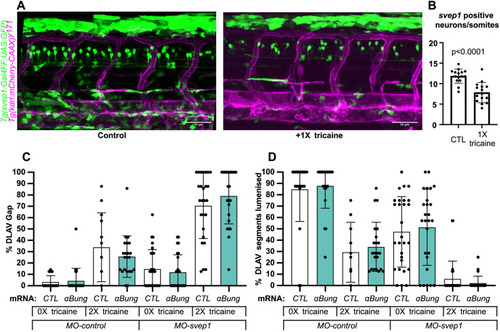
svep1 is expressed in neurons in the neural tube. (A) Representative images of 48 hpf Tg(svep1:Gal4FF; UAS:eGFP); Tg(kdrl:mcherry-CAAX)y171 embryos with or without treatment with 1× (0.0168%) tricaine from 30 to 48 hpf. (B) Quantification of average numbers of Tg(svep1:Gal4FF; UAS:eGFP)-positive neurons in the neural tube area of 48 hpf embryos with or without treatment with 1× (0.0168%) tricaine from 30 to 48 hpf (N=3, n=15 controls, n=16 treated). (C) Bilateral quantifications of the percentage of gaps in the DLAV at 48 hpf in embryos injected with control mRNA (GFP, 50 pg) or alpha-bungarotoxin (αBung) mRNA, MO-CTL (5 ng) or MO-svep1 (5 ng), and treated with 0× or 2× tricaine from 30-48 hpf. (N=2, n=24-28). (D) Bilateral quantifications of the percentage of lumenised segments in the DLAV at 48 hpf in embryos injected with control mRNA (GFP, 50 pg) or αBung mRNA, MO-CTL (5 ng) or MO-svep1 (5 ng) and treated with 0× or 2× tricaine from 30 to 48 hpf (N=2, n=24-28). Data are mean±s.d. Mann–Whitney test. Scale bars: 50 μm (A).
|
