FIGURE

Figure 3.

ID
ZDB-FIG-220404-23
Publication
Wiggin et al., 2022 - V3 Interneurons are Active and Recruit Spinal Motor Neurons During In Vivo Fictive Swimming in Larval Zebrafish
Other Figures
All Figure Page
Back to All Figure Page
Figure 3.

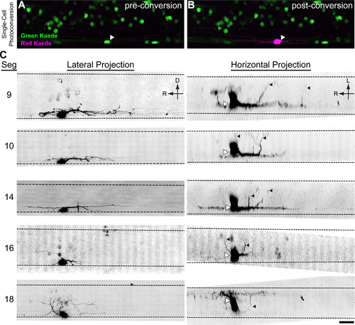
V3-INs are descending, bilaterally projecting cells. Kaede was expressed in vglut2a-expressing neurons in Tg(vglut2a:Gal4ff)nns20;Tg(UAS:Kaede)s1999t double transgenic larvae (A). A single V3-IN was exposed to UV laser light, converting the Kaede protein from green fluorescence to red (B). C, Inverted contrast projections of Kaede-red fluorescence in five neurons. Each row of images is a neuron shown in a lateral z-projection (left) and horizontal z-projection (right). On the horizontal projections, the center of the cell body is indicated with white triangles to account for optical bloom and contralateral projections are indicated with black triangles. Scale bar: 20 μm.

Expression Data

Expression Detail
Antibody Labeling
Phenotype Data

Phenotype Detail
Acknowledgments
This image is the copyrighted work of the attributed author or publisher, and ZFIN has permission only to display this image to its users. Additional permissions should be obtained from the applicable author or publisher of the image. Full text @ eNeuro