IMAGE

Figure 3.

ID
ZDB-IMAGE-220404-23
Source
Figures for Wiggin et al., 2022
Image
Figure Caption

Figure 3.

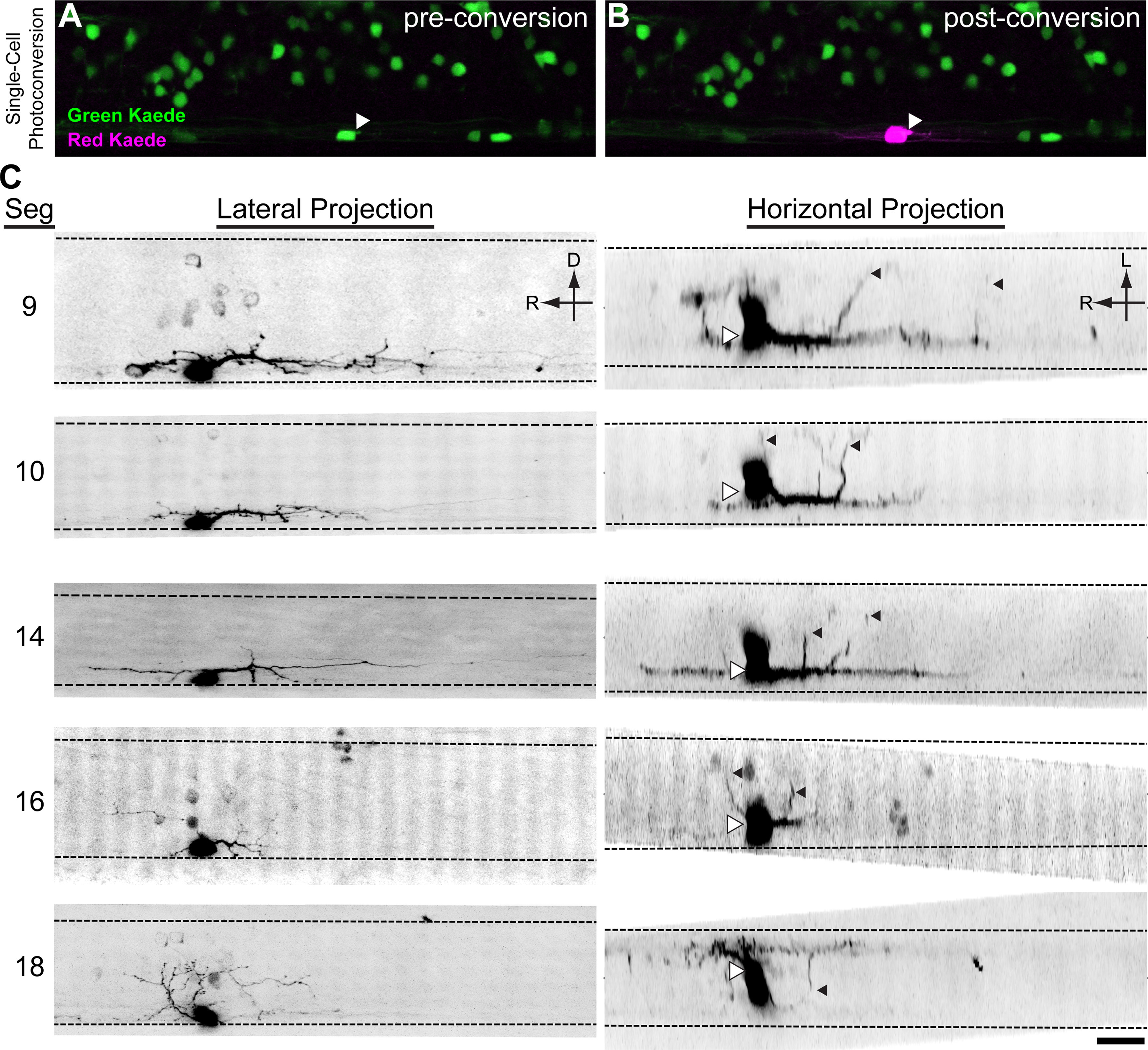
V3-INs are descending, bilaterally projecting cells. Kaede was expressed in vglut2a-expressing neurons in Tg(vglut2a:Gal4ff)nns20;Tg(UAS:Kaede)s1999t double transgenic larvae (A). A single V3-IN was exposed to UV laser light, converting the Kaede protein from green fluorescence to red (B). C, Inverted contrast projections of Kaede-red fluorescence in five neurons. Each row of images is a neuron shown in a lateral z-projection (left) and horizontal z-projection (right). On the horizontal projections, the center of the cell body is indicated with white triangles to account for optical bloom and contralateral projections are indicated with black triangles. Scale bar: 20 μm.

Acknowledgments
This image is the copyrighted work of the attributed author or publisher, and ZFIN has permission only to display this image to its users. Additional permissions should be obtained from the applicable author or publisher of the image. Full text @ eNeuro