|
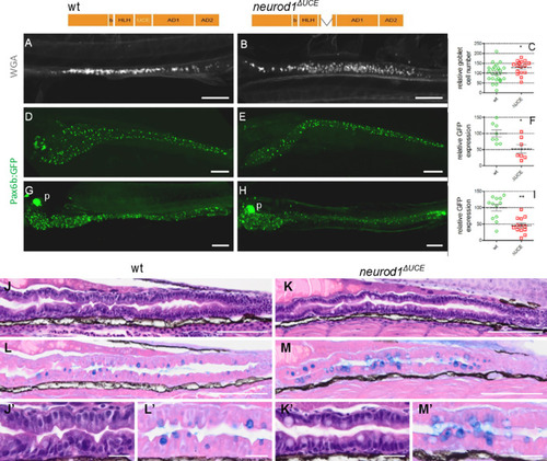
<italic toggle='yes'>neurod1</italic><sup><italic toggle='yes'>ΔUCE</italic></sup> homozygous mutants display an increase of the goblet cells concomitant with a decrease of EECs.Top: Schematic representation of Neurod1 and of Neurod1ΔUCE mutant, harbouring an 81-bp deletion spanning the 19aa of the UCE and 3 and 5 aa upstream and downstream of the UCE, respectively. A-C: Immunodetection of goblet cells labelled with a rhodamine dextran-conjugated wheat germ agglutinin (WGA) that interacts with the N-acetylglucosamine of the mucus. Quantification was done by counting the goblet cells under a fluorescent stereomicroscope. D-I: Immunodetection of GFP performed on pax6b:GFP transgenic embryos at 5 dpf (D-F) or 4 dpf (G-I). The location of the pancreas (p) is indicated on G and H. Quantification of the relative GFP expression was performed by quantifying the volume occupied by the GFP cells using the Imaris software. Asterisks indicate that the difference between the cell number in wt controls and neurod1ΔUCE mutants is statistically significant using the Mann-Whitney U-test (**: P <0.01; *: P <0.05). Views are lateral (A-B) or ventral (D-H) with the anterior part to the left and represent confocal projection images. Scale bars: 100μm. J-M: Subsequent 5 μm microtome sections of 5 dpf wt (J, L) or Neurod1ΔUCE mutant (K,M) larvae stained with haematoxylin/eosin (J-K) or with eosin/alcian blue (L-M). Scale Bars: 100 μm. J’-M’: enlarged view of J-M: Scale Bars: 20 μm.
|