
Fig. 9.
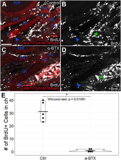
Later paralysis leads to reductions in chondrocyte proliferation. (A-D) Anti-BrdU antibody staining of wild-type (A,B) and α-BTX-injected paralyzed (C,D) sox10:lyn-tdTomato transgenic embryos at 120 hpf. BrdU staining is absent from cartilages, but not from other tissues in α-BTX-injected animals. Blue arrowheads indicate the ventral aspect of the ch cartilage where chondrocyte proliferation appears to concentrate. Green arrowheads indicate proliferation in tissues other than cartilage. All micrographs are ventral view optical slices with anterior to the left. (E) Quantification of BrdU-labeled cells in the ch cartilage. cb, ceratobranchial; D, dorsal; ch, ceratohyal; hs, hyosymplectic pq, palatoquadrate; V, ventral. Scale bar: 50 μm (A-D). |