
Fig. 6
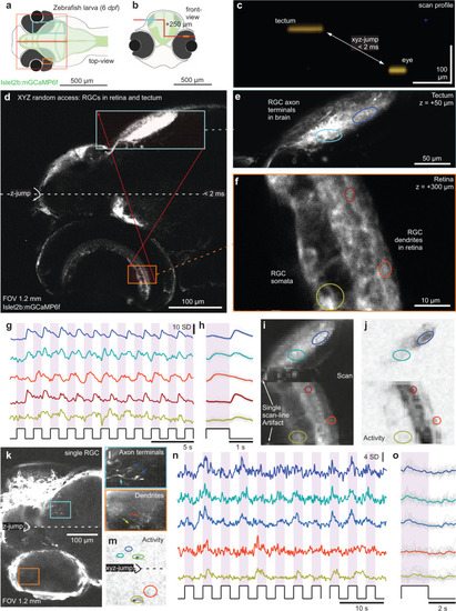
3D random access scanning of the zebrafish eye and brain.
a, b Schematic of zebrafish larva from top (a) and front (b) with scan configurations indicated. c direct x-z visualisation of the scan-profile used in the below. d nTC1 1024 × 1024 px scan across an Islet2b:mGCaMP6f 6 dpf larval zebrafish eye and brain. At the centre of the scan, the axial focus is shifted upwards such that the axonal processes of retinal ganglion cells (RGCs) in the tectum (top) and their somata and dendritic processes in the eye (bottom) can be quasi-simultaneously captured. e, f, 1024 × 1024 px split-plane random access jump between tectum (e) and eye (f) and g–j 2 times 64 × 128 px (15.6 Hz) random access scan of the same scan regions with raw (g) and event-averaged (h) fluorescence traces, mean image (i) and activity-correlation (j, cf. Fig. 3c). The stimulus was a series of full-field broadband flashes of light as indicated. k–o as (d–j), with individual RGCs transiently expressing GCaMP6f under the same promoter. |