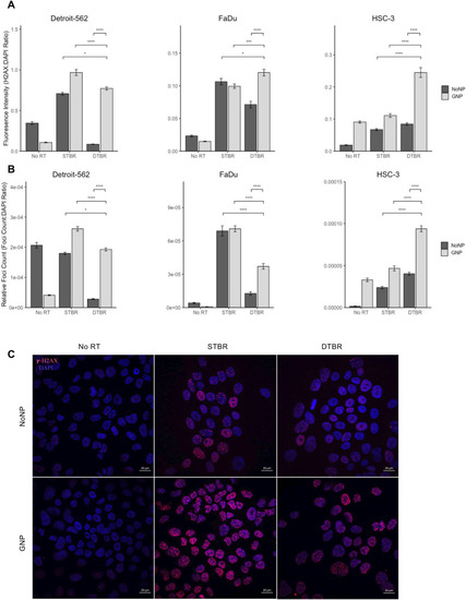
GNP labelled Detroit-562 and HSC-3 HNCCs demonstrate increased fluorescence intensity (FI) of double stranded breaks (DSBs) with DTBR compared to NoNP cells with STBR. Cells were cultured in 6-well plates, irradiated with 8 Gy from the STB or DTB, then fixed 30 min after radiation. Cells were processed for immunohistochemistry (IHC) with γ-H2AX to assess DNA double strand breaks and DAPI nuclear stain. (a) The FI of γ-H2AX foci was determined and measured relative to the FI of DAPI nuclear stain within each nucleus. GNP-labeled Detroit-562 and HSC-3 cells that received DTBR demonstrated increased FI of γ-H2AX foci than No NP cells with STBR (p = 0.049 and p < 0.0001, respectively). With DTBR, Detroit-562, FaDu, and HSC-3 GNP-labeled cells demonstrated greater FI of γ-H2AX foci compared to No NPs (p < 0.0001 for all). GNP-labeled FaDu and HSC-3 cells exhibited increased FI with DTBR compared to STBR (p = 0.001 and p < 0.0001, respectively), but GNP-labeled Detroit-562 cells displayed decreased FI with DTBR compared to STBR (p < 0.0001). (b) The number of γ-H2AX foci was analysed and made relative to the nuclear area. With DTBR, GNP-labeled Detroit-562, FaDu, and HSC-3 cells exhibited significantly greater foci/nucleus than No NP cells (p < 0.0001, p < 0.0001, and p < 0.001, respectively). GNP-labeled Detroit-562 and HSC-3 cells that received DTBR display greater foci/nucleus than No NP cells with STBR (p = 0.0397 and p < 0.0001, respectively), but GNP-labeled FaDu cells with DTBR demonstrated a decrease in foci/nucleus compared to No NP cells with STBR (p < 0.0001). Lastly, GNP-labeled Detroit-562 and FaDu cells with DTBR displayed significantly less foci/nucleus than with STBR (p < 0.0001 for both), but HSC-3 cells exhibited more (p < 0.0001). (c) Representative confocal images of Detroit-562 cells post RT (Zeiss LSM 710 Laser Scanning Confocal microscope at 40 ×). Scale bars = 20 μm. Results are presented as means of [a] Relative fluorescence intensity and [b] Relative Foci count ± standard error of the mean, p* < 0.05, p** < 0.01, p*** < 0.001, p**** < 0.0001. Significance between groups was tested using a standard t-test with Tukey multiple comparison test (n = 3; 4 images/sample). GNP gold nanoparticles, NoNP no nanoparticles, HNCC head and neck cancer cells, STB(R) standard target beam (radiation), DTB(R) diamond target beam (radiation).
|
