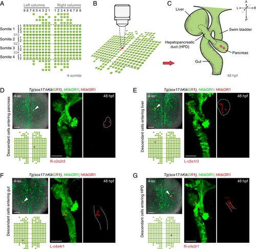
Establishment of single-cell labeling and descendant tracing techniques for endodermal progenitor cells. (A–C) Strategy of single-cell labeling and descendant tracing of early foregut endoderm. At the 4-somite stage, the 273 endodermal cells subjected to single-cell descendant tracing were aligned in a standard map. The dashed lines represent boundaries of somites (A). A single endodermal progenitor cell was labeled by green-to-red photoconversion (B). Then, localizations of its descendants at 48 hpf were detected based on the inheritance of fluorescent retention (C). (D–G) The cells at the positions R-c2s2r2 (D, n = 8), L-c5s1r3 (E, n = 8), L-c4s4r1 (F, n = 7), and R-c4s3r1 (G, n = 8), as indicated in the standard map, were individually labeled by photoconversions. Localizations of their descendant cells at 48 hpf were determined by red fluorescence according to C. Liver and pancreatic buds at 48 hpf stand to the left and right of the gut tube, respectively. (Scale bar, 50 μm.) 4-so, 4-somite stage.
|
