
FIGURE
Figure 1
Figure 1
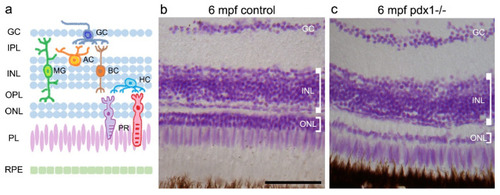
Pathology in the neural retina of pdx1−/− mutants at 6 mpf. (a) Schematic showing the locations of the cell types of the neural retina. Feulgen staining of cryosections of control (b) and pdx1 mutant (c) zebrafish. Scale bar: 50 µm. (GC, ganglion cell layer; IPL, inner plexiform layer; INL, inner nuclear layer; OPL, outer plexiform layer; ONL, outer nuclear layer; PL, photoreceptor layer; RPE, retinal pigment epithelium; AC, amacrine cell; MG, Müller glial cell; BC, bipolar cell; HC, horizontal cell; PR, photoreceptor). |
Expression Data
Expression Detail
Antibody Labeling
Phenotype Data
| Fish: | |
|---|---|
| Observed In: | |
| Stage: | Adult |
Phenotype Detail
Acknowledgments
This image is the copyrighted work of the attributed author or publisher, and
ZFIN has permission only to display this image to its users.
Additional permissions should be obtained from the applicable author or publisher of the image.
Full text @ Cells