
Fig. 2
- ID
- ZDB-FIG-211122-9
- Publication
- Chen et al., 2021 - In vivo volumetric imaging of calcium and glutamate activity at synapses with high spatiotemporal resolution
- Other Figures
- All Figure Page
- Back to All Figure Page
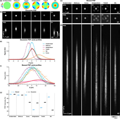
Aberration modes differentially degrade signal and point spread function (PSF) profiles of Gaussian and Bessel foci.
a Zernike modes representing defocus, coma, astigmatism, trefoil, and spherical aberrations (SA), respectively, are introduced to the pupil plane (SLM2). b Lateral (XY) and axial (XZ) PSFs of the Gaussian focus measured using a 0.1-µm-diameter fluorescent bead. c Lateral PSFs of the Bessel focus (upper row) simulated and (lower row) measured using a 0.1-µm-diameter fluorescent bead. d Axial PSF of the Bessel foci measured using a 0.1-µm-diameter fluorescent bead. e, f Axial profiles of the Gaussian and Bessel PSFs, respectively. g Peak signals of Gaussian and Bessel PSFs under different aberration modes. Normalized to the unaberrated PSFs. n = 4 measurements for each aberration mode were acquired from the same bead. Data are presented as dot plots with lines representing mean values. AU arbitrary unit. Source data are available as a source data file. |