
Figure 2
- ID
- ZDB-FIG-211025-49
- Publication
- Paolini et al., 2021 - Mechanosensitive Notch-Dll4 and Klf2-Wnt9 signaling pathways intersect in guiding valvulogenesis in zebrafish
- Other Figures
- All Figure Page
- Back to All Figure Page
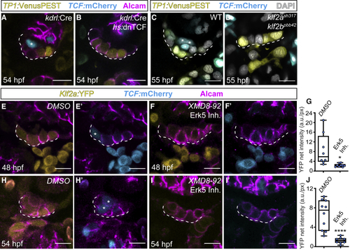
Erk5-Klf2a-Wnt signaling within AVC endocardium is essential for valvulogenesis (A–D) Single confocal z section plane images of the superior AVC endocardium at 54 hpf. (A and B) The endothelial-specific knockdown of canonical Wnt signaling prevents AVC endocardial cells from ingressing into the cardiac jelly. (A) Presence of a TCF-positive ingressed cell when TCF is not blocked (asterisk). Note the presence of TCF-positive cells in the myocardium of treated hearts (B), which confirms the tissue-specificity of this treatment. (C and D) TCF-positive endocardial cells (asterisks in C) are absent in in (E–J) Single confocal z section plane images of the superior AVC endocardium at 54 hpf. (E–G and H–J) Quantification of |
| Genes: | |
|---|---|
| Antibody: | |
| Fish: | |
| Conditions: | |
| Anatomical Terms: | |
| Stage: | Long-pec |
| Fish: | |
|---|---|
| Conditions: | |
| Observed In: | |
| Stage: | Long-pec |