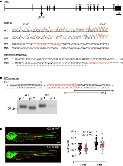
Generation of a CD18 KO zebrafish and analysis of neutrophil count. (A) Upper panel: Schematic of itgb2 gene and target exon 3 of the gRNA1. Middle panel: Sequencing traces and partial genomic sequence of WT and CD18 mutant (mut.). Numbers indicate position within the gene. Red boxes show nucleotides deleted in the mutant. Highlighted is the target sequence of the gRNA in red. Lower panel: Predicted amino acid sequence of mutants aligned to WT sequence of the first 64 amino acids. Identical (*) and altered (red) amino acids are indicated. (B) Upper panel: Location of primer pairs for PCR analysis of WT and CD18 mutant mRNA expression analysis. Reverse (rev) primer 1 binds to WT and CD18 mutant sequence, reverse primer 2 only binds the WT sequence. Forward (for) primer 1 was combined with both reverse primers. Lower panel: Representative image of agarose gel electrophoresis of PCR products of WT and CD18 mutant cDNA with primer pairs (pp) 1 (forward primer 1, reverse primer 1) and 2 (forward primer 1, reverse primer 2), respectively, from zebrafish larvae at 5 dpf. Expected band sizes are 117 bp for pp 1 and 110 bp for pp 2. (C) Left: Exemplary maximum intensity projections of CD18 WT and CD18 KO1 zebrafish larvae at 3 dpf. Endothelial cells are shown in green, neutrophils in red. Scale bars represent 200 µm. Right: Total neutrophil counts in CD18 WT and CD18 KO1 zebrafish larvae at 3 dpf and 5 dpf. Mean ± sem of ≥ 44 individual larvae of ≥ 3 independent experiments. Unpaired t-test.
|
