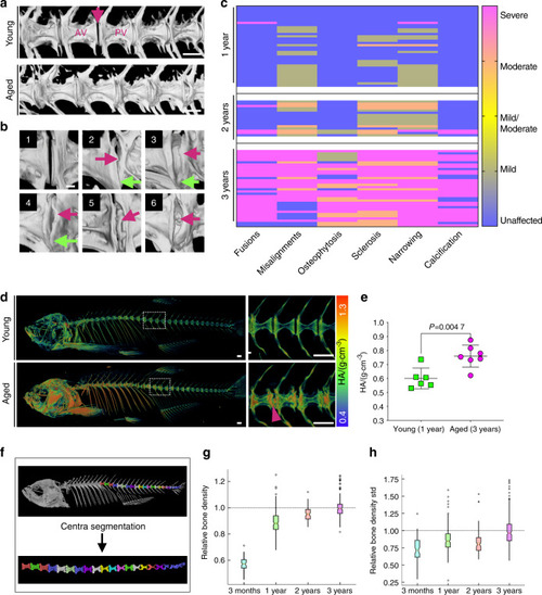
Progressive abnormalities found in aged zebrafish vertebral columns. a 3D rendering from μCT images of young (1 year) and aged (3 years) spines. AV anterior vertebra, PV posterior vertebra, IVD (arrow). Scale bar = 500 μm. b Frequent changes observed at the IVD. 1: normal IVD; 2: osteophytes (pink arrow), vertebral misalignment and IVD narrowing (green arrow); 3: endplate sclerosis; 4: sclerosis (pink arrow) and IVD narrowing (green arrow); 5: sclerosis and fusion (pink arrow); 6: sclerosis and IVD calcification (pink arrow). Scale bar = 100 μm. c Heat map graph showing spinal morphological changes classified by severity during aging (1 year n = 36 (42% females, 58% males), 2 years n = 16 (57% females, 43% males), and 3 years n = 34 (55% females, 45% males)). Average fish standard lengths (measured from tip of the head to the last vertebral column): 1 year = 3.22 cm (0.19 SD), 2 years = 3.41 cm (0.18 SD), and 3 years = 3.46 cm (0.23 SD). d 3D rendering from μCT images of young and aged fish, color coded to show bone mineral density changes. The selected area of the spine (dashed box) is magnified, as shown on the right of the panel. Higher density colocalizes with regions of sclerosis and deformities (arrowhead) in the aged spine. Scale bar = 500 μm. e TMD (tissue mineral density) retrieved from the third thoracic vertebrae in young (1 year) and aged (3 years) fish. Nonparametric, two-tailed, Mann–Whitney test; data are the mean and SD. P values are indicated. f 3D volume rendering from a μCT image of wt fish showing an individual vertebral centrum segmented by computational automation. g Relative bone density from the vertebral centra in aging fish (3 months to 3 years). Average standard lengths: 3 months = 2.6 cm (0.18 SD); 3 years = 3.46 cm (0.23 SD). The notch plot was scaled by the average value from the 3-year centra. h Within-sample standard deviation in bone density. The notch plot was scaled by its average value from the 3-year centra. c, e Generated in Prism 8. g, h Graph was generated in Python
|
