FIGURE

FIGURE 2

ID
ZDB-FIG-210909-5
Publication
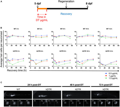
Jimenez et al., 2021 - Vestibular and Auditory Hair Cell Regeneration Following Targeted Ablation of Hair Cells With Diphtheria Toxin in Zebrafish
Other Figures
All Figure Page
Back to All Figure Page
FIGURE 2

Tg(myo6b:hDTR) larval zebrafish show hair cell loss and regeneration in lateral line neuromasts after in vivo DT treatment. (A) Schematic representation of the larval DT exposure treatment. (B) Wild-type (WT) and Tg(myo6b:hDTR) (hDTR) larvae were exposed to three concentrations of DT (0.5, 1, and 1.5 μg/mL) for various durations (3, 6, 8, 10, and 12 h). Neuromast viability was monitored daily by YO-PRO-1 labeling 0 to 72 h post-incubation. Shown is the quantification of YO-PRO-1 labeled hair cells. The values are represented as the mean ± SEM from eight fish. (C) Regeneration time-course showing P1–P4 over three days of recovery. Larvae were exposed to 1 μg/mL of DT for 6 h. Scale bar 100 μm.

Expression Data

Expression Detail
Antibody Labeling
Phenotype Data

Phenotype Detail
Acknowledgments
This image is the copyrighted work of the attributed author or publisher, and ZFIN has permission only to display this image to its users. Additional permissions should be obtained from the applicable author or publisher of the image. Full text @ Front. Cell. Neurosci.