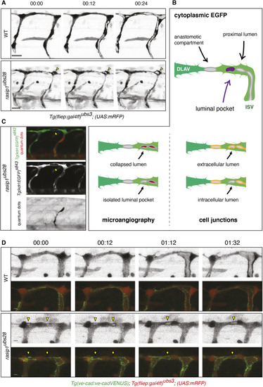
Analysis of ectopic luminal pockets during DLAV formation in rasip1 mutants. (A) Still images of time-lapse movies showing the emergence of ectopic luminal pockets (yellow arrowheads) in rasip1 mutants (Movies 9 and 10). (B) Schematic of possible cellular localizations of ectopic lumens. To differentiate between these possibilities, two types of experiments were performed: microangiography (C) and colocalization of luminal pockets with junctional marker (D). (C) Visualization of ectopic lumens and patent lumens in a rasip1ubs28 embryo (36 hpf). Ectopic luminal pockets are indirectly visualized by the absence of cytoplasmic EGFP (yellow arrowheads) [Tg(kdrl:EGFP)s843]. The patent lumen is marked by microangiography using quantum dots in red (black in bottom panel). Ectopic lumens are not part of the patent vasculature. (D) Still images of time-lapse movies during lumen formation in the DLAV from around 32 hpf onward in wild-type (WT; top) and rasip1ubs28 (bottom) embryos (Movies 11-14). Endothelial cells are labeled with mRFP (grayscale images) and junctions are labeled by VE-cad-Venus (merged images). Yellow arrowheads indicate the ectopic luminal pockets in the rasip1 mutant. Scale bars: 5 μm.
|
