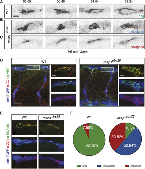
Requirement of Rasip1 for dynamic re-localization of junctional proteins and junctional ring formation during anastomosis. (A-C) Still images of time-lapse movies showing normal junctional patch-to-ring transformation in wild type (WT) (A; Movie 3) and aberrant ring formation in rasip1ubs28 mutants (B,C; Movies 4 and 5). Transgenic embryos expressing a VE-cadherin-Venus fusion protein were imaged, starting at 30 hpf. Scale bar: 5 μm. (D) Immunofluorescence analysis of ZO-1 and VE-cadherin in Tg(kdrl:EGFP)s843 at 32 hpf. rasip1ubs28 mutant shows reticulated junctions between two cells in the DLAV; wild-type embryo forms a cleared apical compartment and a ring-shaped junction. Boxed areas indicate the regions shown at higher magnification to the right. Scale bars: 20 μm (main panels); 5 μm (insets). (E) Immunofluorescence analysis of ZO-1 and Esama in Tg(kdrl:EGFP)s843 at 32 hpf showing a collapsed junction in the rasip 1ubs28 mutant. Scale bars: 5 μm. (F) Quantification of observed junctional phenotypes at 32 hpf. rasip1ubs28 mutants show a significant number of reticulated junctions and collapsed anastomotic rings compared with wild type (WT n=6 embryos, 53 analyzed rings; mutant n=8 embryos, 68 analyzed rings). P<0.0001 (χ2 test).
|
