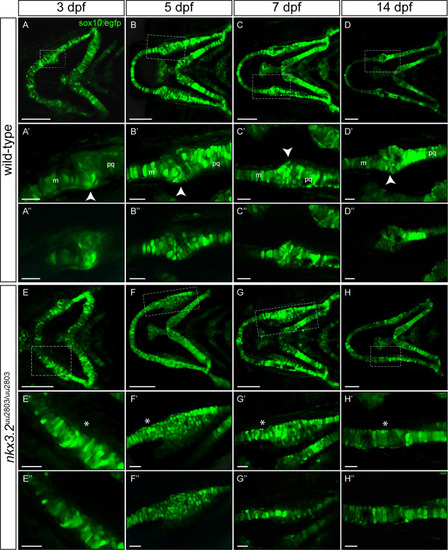
(A-H’) Maximum projections of confocal live imaging Z-stacks acquired from ventral side of wild-type zebrafish head at 3 dpf (A, A’), 5 dpf (B, B’), 7dpf (C, C’), 14 dpf (D, D’) and nkx3.2uu2803/uu2803 zebrafish head at 3 dpf (E, E’), 5 dpf (F, F’), 7 dpf (G, G’), 14 dpf (H, H’) in Tg(sox10:egfp) background. Corresponding single Z-planes are shown in A”–H”. (A-D´´) The jaw joint in wild-type zebrafish (dashed box) is magnified in A’-D”. The retroarticular process is visible from 3 dpf onwards, marked by white arrowhead. Chondrocytes of the anterior Meckel’s cartilage and posterior palatoquadrate align in stacks. Posterior Meckel’s cartilage and anterior palatoquadrate articulate the jaw joint. (E-H”) Fusion of jaw joint articulating elements in nkx3.2 mutants. The fusion site is magnified in (E’-H”) and indicated by asterisks. (E’-F’ and E”-F”) At 3 dpf and 5 dpf nkx3.2 mutants display unorganised and rounded cells in the anterior palatoquadrate, posterior to the jaw joint fusion site (G’, G”) At 7 dpf nkx3.2 mutants display elongated chondrocytes that start to align in stacks. (H’, H”) At 14 dpf nkx3.2 mutants display elongated chondrocytes that are aligned throughout the fused Meckel’s-palatoquadrate element. m—Meckel’s cartilage, pq—palatoquadrate. Scale bars: 100 μm (A-H), 25 μm in (A’-H’ and A”-H”).
|
