
FIGURE
Fig 7
Fig 7
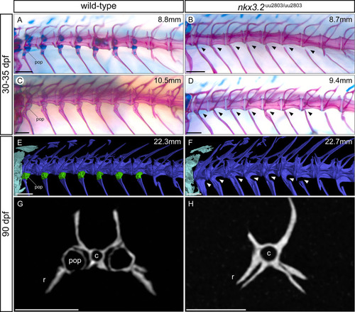
(A-F) Dorsolateral views of rib-bearing vertebrae in 30–35 dpf, and 90 dpf wild-type and nkx3.2 mutant zebrafish. (A-D) Cartilage- and bone- stained juvenile zebrafish, (E, F) μCT models. (G, H) μCT virtual transverse cross-sections of wild-type and nkx3.2 mutant rib-bearing vertebrae at 90 dpf. The measurements in mm refer to standard length (SL). Arrowheads in (B, D, F) indicate the absence of parapophyses on the rib-bearing vertebrae. Parapophyses (pop) are highlighted in green in (E). c–centrum, r–rib. Scale bars: 200μm(A-D), 500μm (E-H). |
Expression Data
Expression Detail
Antibody Labeling
Phenotype Data
| Fish: | |
|---|---|
| Observed In: | |
| Stage Range: | Days 30-44 to Adult |
Phenotype Detail
Acknowledgments
This image is the copyrighted work of the attributed author or publisher, and
ZFIN has permission only to display this image to its users.
Additional permissions should be obtained from the applicable author or publisher of the image.
Full text @ PLoS One