|
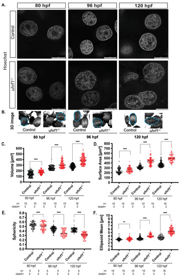
uhrf1 loss increases hepatocyte nuclear size, disrupts nuclear morphology, and increases nucleoli number at 120 hpf. (A). Confocal images of phenotypically WT siblings and uhrf1−/− nuclei stained by Hoechst (in grey). (B). The 3D reconstructed nuclei in the liver of 80, 96, and 120 hpf WT siblings and uhrf1−/− nuclei larvae display changes in size and shape at all time points between controls and mutants. (C–F). Nuclear morphological measurements of control and uhrf1−/− hepatocyte nuclei scored at 80, 96, and 120 hpf for volume (C), surface area (D) for size measurements, and sphericity (E), ellipsoid mean (F) for shape measurements. Each dot represents one cell. All analyses were performed on 2 or 3 clutches, with minimum 2 livers analyzed per clutch. ** p < 0.005, **** p < 0.00005. Magnification: 63×, 4× zoom, Scale bar: 5 μm.
|