
Figure 4
- ID
- ZDB-FIG-210727-39
- Publication
- Okuda et al., 2021 - 3,4-Difluorobenzocurcumin Inhibits Vegfc-Vegfr3-Erk Signalling to Block Developmental Lymphangiogenesis in Zebrafish
- Other Figures
- All Figure Page
- Back to All Figure Page
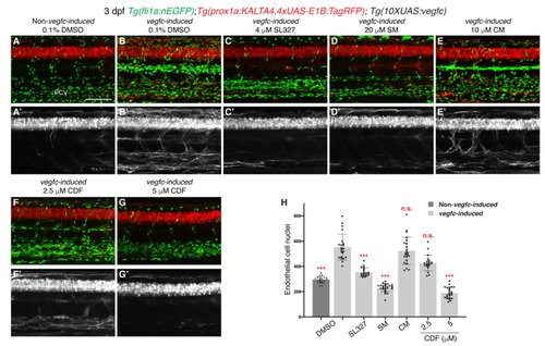
3,4-Difluorobenzocurcumin inhibits pathological phenotypes associated with vegfc overexpression. (A–G’) Lateral confocal images of either a 3 dpf Tg(prox1a:KALTA4,4xUAS-E1B:TagRFP);Tg(fli1a:nEGFP) larva (Non-vegfc-induced) treated with 0.1% DMSO (A,A’), or 3 dpf Tg(prox1a:KALTA4,4xUAS-E1B:TagRFP);Tg(10XUAS:vegfc);Tg(fli1a:nEGFP) larvae (vegfc-induced) treated with either 0.1% DMSO (B,B’), 4 μM SL327 (C,C’), 20 μM sunitinib malate (SM, D,D’), 10 μM curcumin (CM, E,E’), 2,5 μM 3,4-Difluorobenzocurcumin (CDF, F,F’), or 5 μM CDF (G,G’). Pathological vascular phenotypes in vegfc-induced embryos are rescued by CDF treatment. Images (A’–G’) represent the Tg(prox1a:KALTA4,4xUAS-E1B:TagRFP) expression of images (A–G). To avoid the robust prox1a expression in muscle cells, (A’–G’) are maximum projection images of only the z stacks that contain the posterior cardinal vein. Images (B’) (21/22 embryos), (E’) (23/23 embryos) and (F’) (14/20 embryos) show embryos with increased prox1a:KALTA4,4xUAS-E1B:TagRFP expression in venous endothelial cells. This pathological phenotype is rescued in images (C’) (20/20 embryos), (D’) (27/27 embryos) and (G’) (21/24 embryos). (H) Quantification of fli1a:EGFP-positive ECs across 4.5 somites in either 3 dpf non-vegfc-induced treated with 0.1% DMSO (n = 21 embryos) or 3 dpf vegfc-induced larvae treated with either 0.1% DMSO (n = 22 embryos), 4 μM SL327 (n = 20 embryos), 20 μM SM (n = 27 embryos), 10 μM CM (n = 23 embryos), or CDF at 2.5 μM (n = 20 embryos) or 5 μM (n = 24 embryos). PCV: posterior cardinal vein. Statistical test: Kruskal-Wallis test was conducted for graph H. p ≤ 0.001 (***) and n.s. indicates not significant. Scale bar: 100 μm. |