
FIGURE
FIGURE 4
- ID
- ZDB-FIG-210718-31
- Publication
- Shimizu et al., 2021 - Differential Regenerative Capacity of the Optic Tectum of Adult Medaka and Zebrafish
- Other Figures
- All Figure Page
- Back to All Figure Page
FIGURE 4
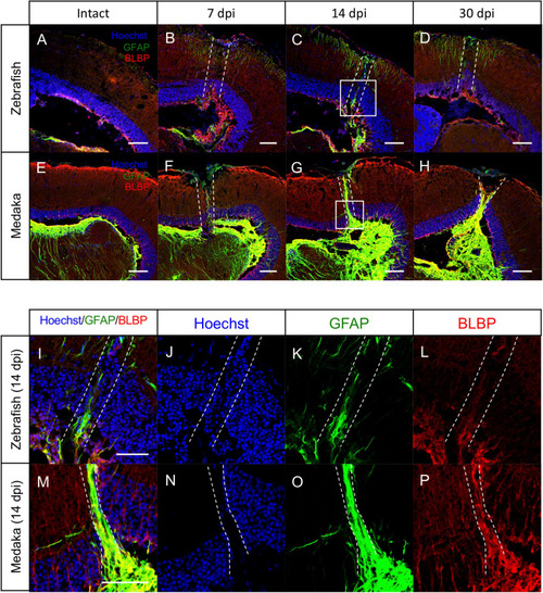
Persistent glial scar-like structure is observed in the injured medaka tectum. |
Expression Data
Expression Detail
Antibody Labeling
Phenotype Data
Phenotype Detail
Acknowledgments
This image is the copyrighted work of the attributed author or publisher, and
ZFIN has permission only to display this image to its users.
Additional permissions should be obtained from the applicable author or publisher of the image.
Full text @ Front Cell Dev Biol