
FIGURE
FIGURE 8
- ID
- ZDB-FIG-210717-27
- Publication
- Afonin et al., 2021 - In Vivo Behavior of the Antibacterial Peptide Cyclo[RRRWFW], Explored Using a 3-Hydroxychromone-Derived Fluorescent Amino Acid
- Other Figures
- All Figure Page
- Back to All Figure Page
FIGURE 8
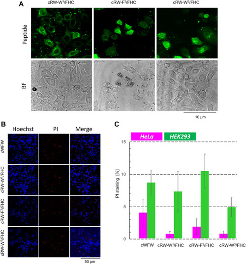
Cytotoxicity evaluation of cWFW and its fluorescent analogues by fluorescence microscopy. |
Expression Data
Expression Detail
Antibody Labeling
Phenotype Data
Phenotype Detail
Acknowledgments
This image is the copyrighted work of the attributed author or publisher, and
ZFIN has permission only to display this image to its users.
Additional permissions should be obtained from the applicable author or publisher of the image.
Full text @ Front Chem