IMAGE

FIGURE 8

ID
ZDB-IMAGE-210717-27
Source
Figures for Afonin et al., 2021
Image
Figure Caption

FIGURE 8

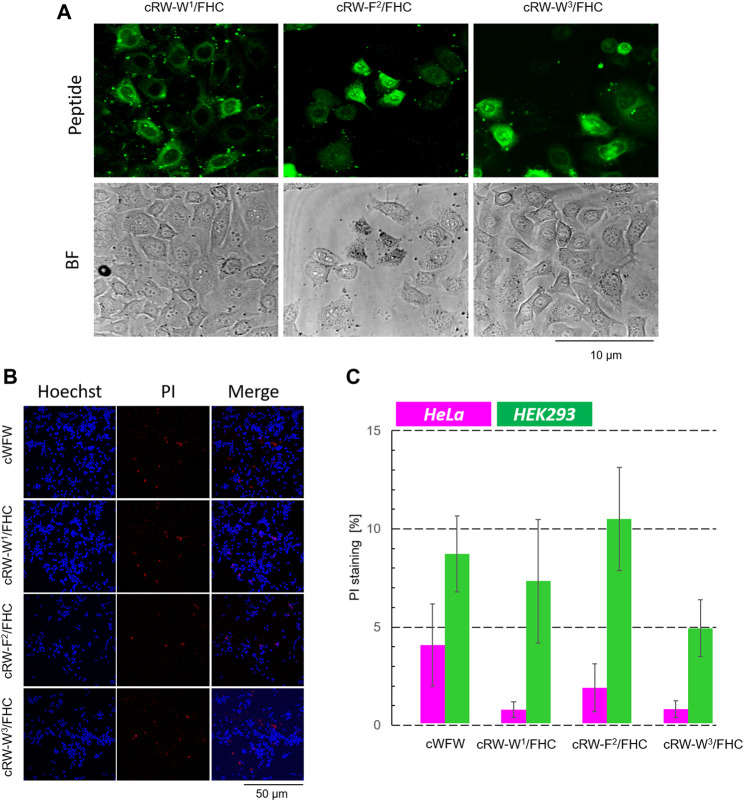
Cytotoxicity evaluation of cWFW and its fluorescent analogues by fluorescence microscopy. (A) First, at higher magnification (63× objective), the cells were imaged to monitor cell entry of the peptides; HeLa cells with 20 µM peptide imaging after 1 h of incubation are shown for illustration. (B) After imaging (2.5 h after peptide addition), Hoechst/PI co-staining is applied and imaged at 20× magnification, exemplified here by experiments with HEK293 cells; (C) Total/dead cell counting results for all peptides at 20 µM concentration co-incubated with both cell types.

Acknowledgments
This image is the copyrighted work of the attributed author or publisher, and ZFIN has permission only to display this image to its users. Additional permissions should be obtained from the applicable author or publisher of the image. Full text @ Front Chem