
Figure 3
- ID
- ZDB-FIG-210607-30
- Publication
- Li et al., 2021 - Abortive intussusceptive angiogenesis causes multi-cavernous vascular malformations
- Other Figures
-
- Figure 1
- Figure 1 - figure supplement 1
- Figure 1 - figure supplement 2
- Figure 2
- Figure 3
- Figure 3 - figure supplement 1
- Figure 4
- Figure 4 - figure supplement 1
- Figure 4 - figure supplement 2
- Figure 5
- Figure 5 - figure supplement 1
- Figure 6
- Figure 6 - figure supplement 1
- Figure 7
- Figure 7 - figure supplement 1
- Figure 7 - figure supplement 2
- All Figure Page
- Back to All Figure Page
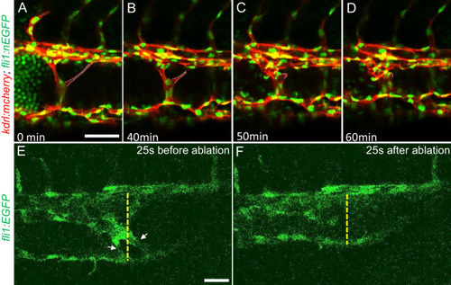
(A through D) Time lapse images reveal spontaneous retraction of an intravascular pillar leading to re-entry of blood cells into circulation and reduced dilation of the caudal vein. Endothelial cells were labeled by mCherry, and their nucleus and some red blood cells were labeled by EGFP in the Tg(fli1:nEGFP)y7;Tg(kdrl:mcherryras)s896 embryos. The retracted pillar is outlined by dotted lines for emphasis. Note that pillar retraction and vessel dilation were temporally correlated. (E and F) Laser ablation of pillar reduced caudal venous plexus (CVP) diameter. The diameter of the dilated vein (E) was reduced after ablation (F). Note the pillars indicated by arrows in (E) are gone after ablation in (F). Dashed line indicates the diameter of the vein before and after ablation. Scale bar: 50 µm. |