
Fig. 5
- ID
- ZDB-FIG-210426-28
- Publication
- Atieh et al., 2020 - Pulsatile contractions promote apoptotic cell extrusion in epithelial tissues
- Other Figures
- All Figure Page
- Back to All Figure Page
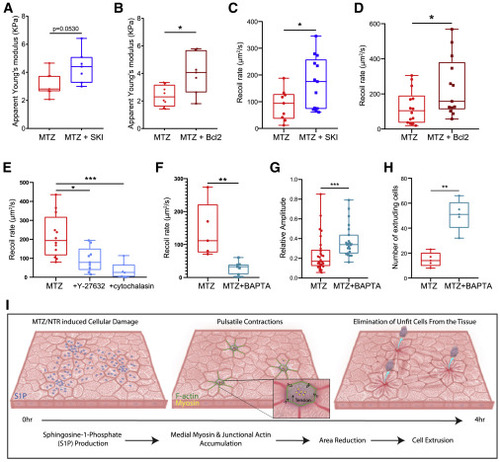
Caspase activation and S1P phosphorylation alter the mechanical state of the tissue following damage (A) Quantification of the apparent stiffness of epithelia of larvae treated with MTZ versus MTZ+SKI. (B) Quantification of the apparent stiffness of epithelia in NTR- versus NTR+Bcl2-overexpressing larvae treated with MTZ. (C) Quantification of the recoil rate in MTZ- versus MTZ+SKI-treated larvae. (D) Quantification of the recoil rate in NTR- versus NTR+Bcl2-overexpressing larvae treated with MTZ. (E) Quantification of the recoil rate in MTZ-, MTZ+Y-27632-, or MTZ+cytochalasin-treated larvae. (F) Quantification of the recoil rate in MTZ- versus MTZ+BAPTA-treated larvae. (G) Quantification of the relative amplitude of pulses in MTZ- versus MTZ+BAPTA-treated larvae. See also Video S6. (H) Quantification of the number of extruding cells in MTZ- versus MTZ+BAPTA-treated larvae. See also Video S6. (I) Upon MTZ treatment, damaged epithelial cells get enriched in S1P. Damage and S1P enrichment lead to decrease in tension within the tissue and induce pulsatile contractions through medio-apical myosin coalescence. Cells that pulse with higher dynamics preferentially extrude. |