
Fig. 6
- ID
- ZDB-FIG-210413-46
- Publication
- Dietrich et al., 2021 - Skeletal biology and disease modeling in zebrafish
- Other Figures
- All Figure Page
- Back to All Figure Page
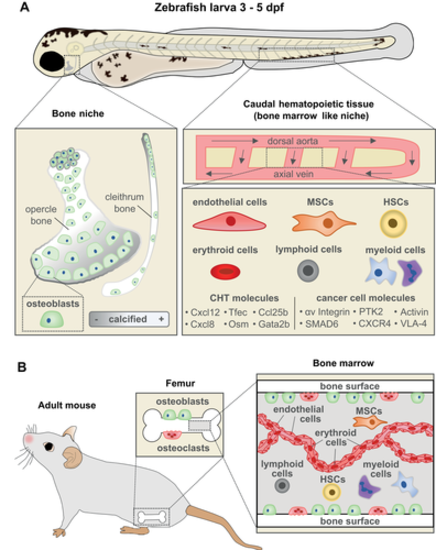
The CHT as an attractive site to study bone metastasis (A) Zebrafish larvae at 3 to 5 dpf contain developing bone (opercle and cleithrum) and CHT which acts as a BM‐like niche. (Left) The opercle and cleithrum are among the first dermal bones to develop and can be imaged in live larvae due to their lateral, superficial positioning. (Right) The CHT is located laterally to the dorsal aorta in the posterior region of the larval zebrafish and is the site of early hematopoiesis, akin to mammalian BM. The cellular components of the CHT include endothelial cells, MSCs, HSCs, myeloid cells such as macrophages and neutrophils, lymphoid cells, and erythroid cells. Molecular components of the CHT listed have been previously shown to play a role in bone metastasis in mammalian models. Metastasis‐related factors expressed in cancer cells uncovered in this model are shown on the right. (B) Illustration of the BM niche in a mouse long bone containing similar cell types as the CHT. dpf = days post fertilization; MSCs = mesenchymal stromal cells; HSCs = hematopoietic stem cells. |