
Fig. 3
- ID
- ZDB-FIG-210324-3
- Publication
- Ye et al., 2020 - Enteroendocrine cells sense bacterial tryptophan catabolites to activate enteric and vagal neuronal pathways
- Other Figures
- All Figure Page
- Back to All Figure Page
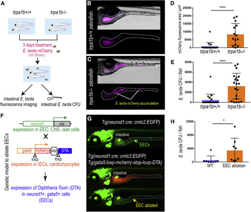
Figure 3. Activation of EEC Trpa1 signaling facilitates enteric E. tarda clearance (A) Schematic of zebrafish E. tarda treatment. (B and C) Representative image of trpa1b+/+ (B) or trpa1b−/− (C) zebrafish treated with E. tarda expressing mCherry (E. tarda mCherry). (D) Quantification of E. tarda mCherry fluorescence in trpa1b+/+ or trpa1b−/− zebrafish intestine. (E) Quantification of intestinal E. tarda CFU in trpa1b+/+ or trpa1b−/− zebrafish. (F) Schematic of a genetic model in which EECs are ablated via Cre-induced Diphtheria Toxin (DTA) expression. (G) Representative image of Tg(neurod1:cre; cmlc2:EGFP) and Tg(neurod1:cre; cmlc2:EGFP); TgBAC(gata5:RSD) with EECs that are labeled by Tg(neurod1:EGFP). (H) Quantification of intestinal E. tarda CFU in WT or EEC-ablated zebrafish. Student’s t test was used in (D, E, and H). ∗p < 0.05; ∗∗∗∗p < 0.0001. |