
Fig. 4
- ID
- ZDB-FIG-210316-30
- Publication
- Yu et al., 2020 - Taste buds are not derived from neural crest in mouse, chicken, and zebrafish
- Other Figures
- All Figure Page
- Back to All Figure Page
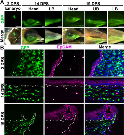
Fig. 4. Distribution of GFP+ neural fold-derived cells in the craniofacial regions ispilateral to the surgery side. A: Photomicrographs of a chimeric embryo at 2 DPS, side view of heads and dorsal view of the lower beak (LB) at 14 and 19 DPS, and upper beak (UP) at 19 DPS. Top panel: fluorescent images to show the GFP signals; Bottom panel: merged fluorescent and bright-field images. B: Single-plane laser scanning confocal images of sagittal sections of pharyngeal arch of chimeric embryo at 2 DPS and base of oral cavities of chimeric embryos at 14 and 19 DPS. Sections were immunoreacted for the epithelial cell marker EpCAM (magenta). White dashed lines demarcate the epithelium from the underlying mesenchyme. Scale bars: 2 mm in A; 20 μm in B. |
Reprinted from Developmental Biology, 471, Yu, W., Wang, Z., Marshall, B., Yoshida, Y., Patel, R., Cui, X., Ball, R., Yin, L., Kawabata, F., Tabata, S., Chen, W., Kelsh, R.N., Lauderdale, J.D., Liu, H.X., Taste buds are not derived from neural crest in mouse, chicken, and zebrafish, 76-88, Copyright (2020) with permission from Elsevier. Full text @ Dev. Biol.