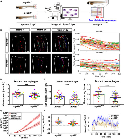
Quantification of distant macrophages behavior in wounded myd88 mutant and sibling control larvae. (A) Experimental scheme. Myd88+/+ and myd88– /– larvae were wounded at 3 dpf. The red dashed line shows the site of wounding. Macrophages of wounded zebrafish larvae were tracked for 2 h and images were taken every 1 min by using CLSM. For cell tracking analysis, cells localized outside an area of 200 μm from the wounding edge toward the body trunk were counted as distant cells. Blue dashed box shows the area where distant macrophages were tracked. (B) Representative images of distant macrophage tracks in the wounded tail fin of 3 dpf myd88+/+ or myd88– /– larvae at frame 1, frame 60 and frame 120. Time interval between two successive frames is 1 min. Each color track represents an individual macrophage. Cell tracking movies are shown in Supplementary Movies S15, S16). Scale bar: 50 μm. (C) Distance to the wound. Black dash line represents average distance to the wound. Each color line represents one cell. (D–I) Quantification of distant macrophage tracks. In (D,F,H), each color indicates a different larva. There was a significant difference between the groups in terms of mean speed (D), net displacement (E), meandering index (F), MSD (red) and fitted MSD (black) (G) and mean VAP(H) of macrophages. Statistical analyses were done with 9 and 8 fish, respectively, for each group. The shaded regions in MSD (G) and mean VAP over time (I) indicate standard error of the mean. An unpaired, two-tailed t-test was used to assess significance (ns, non-significance, ∗∗P < 0.01, ∗∗∗P < 0.001, ****P < 0.0001) and data are shown as mean ± SD. Sample size (n): 50, 44 (D–F,H).
|
