|
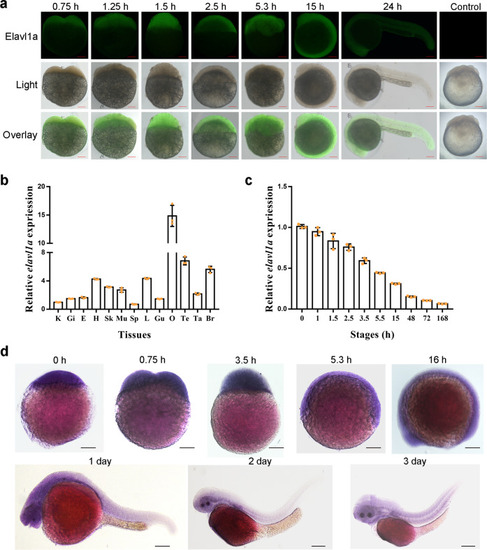
Distribution of zebrafish ELAVL1a in the cells of different developmental stage embryos and expression patterns of zebrafish <italic>elavl1a</italic> in the different tissues as well as at the different developmental stages.a Immunohistochemical localization of ELAVL1a in the different developmental stage: 2-cell stage (about 0.75 h); 8-cell stage (~1.25 h); 16-cell stage (~1.5 h); 256-cell stage (~2.5 h); 50% epiboly stage (~5.3 h); 10-somite stage (~15 h); 24-h post-fertilization (24-hpf); Control: 16-cell embryo incubated with mouse pre-immune serum as control. Scale bars represent 100 μm. b Expression profiles of zebrafish elavl1a in the different tissues including kidney (K), gill (Gi), eye (E), heart (H), skin (Sk), muscle (Mu), spleen (Sp), liver (L), gut (Gu), ovary (O), testis (Te), tail (Ta), and brain (Br). c Expression profiles of zebrafish elavl1a at the different developmental stages including zygote (0 h), 4-cell stage (about 1 h), 16-cell stage (~1.5 h), 256-cell stage (~2.5 h), high blastula stage (~3.5 h), 50% epiboly stage (~5.5 h) embryos, 10-somite stage (~15 h), 2-day post-fertilization (2-dpf), 3-dpf, and 7-dpf larvae. β-actin was chosen as the internal control for normalization. Relative expression data were calculated by the method of 2−∆∆Ct. The vertical bars represent the mean ± SD (n = 3). The data are from three independent experiments performed in triplicate. d Expression of elavl1a during early development detected by WISH. Stages of embryonic development: newly fertilized egg (0 h); 2-cell stage embryo (~0.75 h); high blastula stage embryo (~3.5 h); 50% epiboly stage embryo (~5.3 h); 14-somite larvae (16 h); 1-day-old larvae; 2-day-old larvae; 3-day-old larvae. The color of the positive signal is purple.
|