|
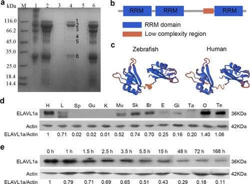
Identification of ELAVL1a as an LTA-binding protein.a SDS-PAGE of the proteins isolated from the embryos extracts of zebrafish on LTA-conjugated Sepharose CL-4B affinity resin. Lane M, marker; lane 1, embryos extracts; lane 2, effluent fractions after Tris-HCl wash; lane 3, effluent fractions after throughout elution; lane 4, effluent fractions containing the absorbed proteins; lane 5, effluent fractions from LTA-conjugated Sepharose CL-4B affinity resin with no embryos extracts loaded; lane 6, effluent fractions from Sepharose CL-4B affinity resin with no conjugated LTA. b Domain structure of ELAVL1a predicted by the SMART program. c 3D structures of the ELAVL1a generated by SWISS-MODEL online software using human ELAVL1 (PDB code: 4egl.1.A) as the model. d Western blotting of ELAVL1a in different tissues including heart (H), liver (L), spleen (Sp), gut (Gu), kidney (K), muscle (Mu), skin (Sk), brain (Br), eye (E), gill (Gi), ovary (O), testis (Te) and tail (Ta). e Western blotting of ELAVL1a at the different developmental stages including zygote (0 h), 4-cell stage (about 1 h), 16-cell stage (~1.5 h), 256-cell stage (~2.5 h), high blastula stage (~3.5 h), 50% epiboly stage (~5.5 h), 10-somite stage (~15 h), 2-day post-fertilization (2-dpf), 3-dpf, and 7-dpf. The values of the quantification of Western blotting were given as well. Data are presented as the means ± SD.
|