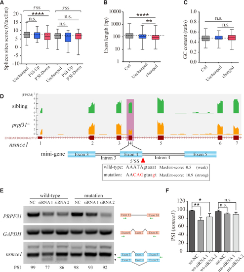
Prpf31 deletion is more likely to cause the skipping of exons with shorter length and weaker 5′ splicing site. (A) The lengths of severely skipped exons between mutants and siblings (‘Changed’ group) were shorter when compared with the exons with no significant difference in PSI values (‘Unchanged’ group) or all the RefSeq annotated exons (‘Ctrl’ group). (B) No significant difference in the GC content of exons between the ‘Changed’ and ‘Unchanged’ or ‘Ctrl’ groups. (C) The splice strength of 5′SS but not 3′SS were weaker in the ‘PSI-Down’ group when compared with the exons in ‘Unchanged’ or ‘PSI-Up’ groups. ‘PSI-Up’, exons with increased PSI values in ‘Changed’ group. ‘PSI-Down’, exons with decreased PSI values in ‘Changed’ group. (D) The skipping of exon 4 in nsmce1 was shown based on the RNA-seq data. The splice strength of this 5′SS was much weaker than the average score (8.4) of ‘Unchanged’ group. Several nucleotides (marked in red) were mutated to enhance the splice strength. (E) Two minigenes containing the weak and strong 5′SS were constructed and tested in HEK293 cells. Knockdown of PRPF31 significantly suppressed the splicing of the wild-type but not the enhanced form of nsmce1-minigene. (F) Quantitative analysis of the PSI values from three independent experiments.
|
