|
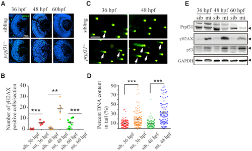
Accumulation of DNA damage in prpf31 mutant retinas. (A) Immunofluorescence analysis using the anti-γH2AX antibody in siblings and prpf31−/− retinas at 36, 48, and 60 hpf. Scale bar, 50 μm. (B) Quantitative analysis of the γH2AX positive cells shown in (A). (C) Alkaline comet assay showed increased DNA damage in prpf31−/− zebrafish at 36 and 48 hpf. Scale bar, 10 μm. White arrows showed DNA with single or double strand breaks. (D) Quantitative results of 100 cells from 6 embryos in each group are shown. White arrows indicate DNA damaged cells. (E) The protein levels of Prpf31, γH2AX and p53 in siblings and prpf31−/− zebrafish at 36, 48 and 60 hpf were detected by western blot. GAPDH was used to normalize protein loading. The black arrows indicated the corresponding protein bands.
|