
Figure 2
- ID
- ZDB-FIG-210301-35
- Publication
- Yao et al., 2021 - Pathways Regulating Establishment and Maintenance of Cardiac Chamber Identity in Zebrafish
- Other Figures
- All Figure Page
- Back to All Figure Page
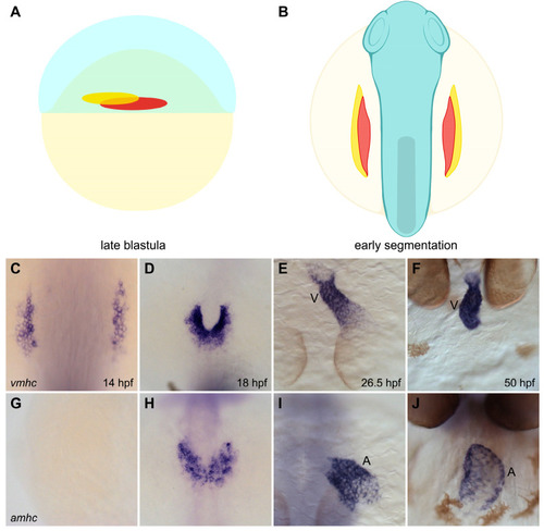
Spatial organization of ventricular and atrial myocardial lineages in the zebrafish embryo. (A,B) Cartoons illustrate the locations of the territories containing ventricular (red) and atrial (yellow) myocardial progenitor cells in the early embryo. Lateral view of the late blastula (A) shows that the regions containing ventricular and atrial myocardial progenitors are spatially organized prior to gastrulation [34]. Dorsal view of the gastrula (B) shows the ALPM regions that contain ventricular and atrial myocardial precursors during early segmentation stages [28]. (C–J) In situ hybridization depicts the expression patterns of vmhc (C–E, dorsal views; F, frontal view) and amhc (G–I, dorsal views; J, frontal view), indicating the relative positions of ventricular and atrial cardiomyocytes as they differentiate and form the heart. Ventricular cardiomyocytes initiate vmhc expression around 14 hpf (C), whereas atrial cardiomyocytes initiate amhc expression around 18 hpf (H) [38,39]; at these stages, ventricular cardiomyocytes are located more medially than atrial cardiomyocytes (D,H). Ventricular and atrial cardiomyocytes go on to occupy separate portions of the heart tube (E,I); later, the ventricular and atrial chambers become morphologically distinct (F,J). V, ventricle, A, atrium. Images adapted from [27]; illustrations by Jessyka T. Diaz. |