FIGURE

Figure 1—figure supplement 1.

ID
ZDB-FIG-210301-134
Publication
Xiong et al., 2021 - In vivo proteomic mapping through GFP-directed proximity-dependent biotin labelling in zebrafish
Other Figures
All Figure Page
Back to All Figure Page
Figure 1—figure supplement 1.

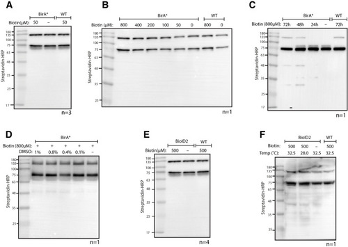
Testing BioID and BioID2 in transgenic zebrafish.

(A) Streptavidin-HRP blot showing biotinylated proteins in BioID (BirA*) transgenic zebrafish embryos. Zebrafish embryos were incubated with 50 µM biotin for 18 hr prior to protein extraction and western blotting. (B, C and D) Streptavidin-HRP blots showing biotinylated proteins in zebrafish embryos with (B) increased biotin concentration, (C) increased biotin incubation time and (D) addition of DMSO. (E and F) Streptavidin-HRP blots showing biotinylated proteins in BioID2 transgenic zebrafish embryos under (E) standard incubation temperature (28°C) and (F) elevated temperature (32.5°C) during biotin incubation. Each sample is a pool of 30 embryos at 3 dpf. The two prominent protein bands present in all samples were endogenous biotinylated proteins.

Expression Data

Expression Detail
Antibody Labeling
Phenotype Data

Phenotype Detail
Acknowledgments
This image is the copyrighted work of the attributed author or publisher, and ZFIN has permission only to display this image to its users. Additional permissions should be obtained from the applicable author or publisher of the image. Full text @ Elife