|
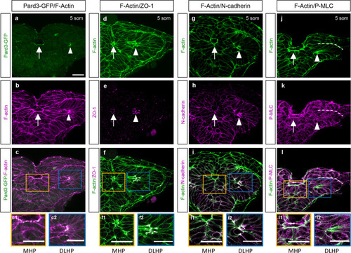
Molecular characterization of the MHP.a–l Transverse sections through the ANP at the 5 som stage. Embryos double-labeled with Pard3-GFP (green) and phalloidin (F-actin, magenta) (a–c2); ZO-1 (magenta) and F-actin (green) (d–f2); N-cadherin (magenta) and F-actin (green) (g–i2); and with anti-P-MLC (magenta) and F-actin (green) (j–l2). c, f, i, l Magenta and green channel overlay. Insets show higher magnification images of the MHP (yellow boxes, c1, f1, i1, l1) and DLHP (blue box, c2, f2, i2, l2). Annotations: arrow = MHP; arrowhead = DLHP; dotted line in j–l = interface between the neuroectoderm and non-neural ectodermlayers of the neural folds; MHP medial hingepoint, DLHP dorsolateral hingepoint. Scale bars: 25 μm.
|