|
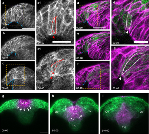
Dynamics of neural fold formation and MHP intercalation.a–f Time-lapse movie frames of the ANP, from a transverse view. a–c Still frames of an embryo expressing membrane Kaede (mKaede). d–f Still frames of a Tg[emx3:YFP] embryo expressing membrane RFP (mRFP, pseudo labeled magenta) and YFP (green). Yellow boxes in a, c, d, and f identify magnified areas in a1, c1, d1, and f1. g–i Still frames of an embryo expressing mKaede (green), in which the MHP cells have been photoconverted (magenta) to follow their fate. MHP medial hingepoint, OV optic vesicles, hyp hypothalamus. Annotations: blue dotted line: outlines the basal side of the neural folds; red and white lines identify individual neural fold cells; arrowhead: indicate narrowing surface in neural folds cells; arrows: show direction of intercalation of MHP cells into the eye field; dotted white line: separation of the superficial and deep layers. Scale bars: 50 μm in a, d and g; 25 μm in a1 and d1.
|