
Figure 5
- ID
- ZDB-FIG-210128-139
- Publication
- Zhou et al., 2021 - Development of a Mucus Gland Bioreactor in Loach Paramisgurnus dabryanus
- Other Figures
- All Figure Page
- Back to All Figure Page
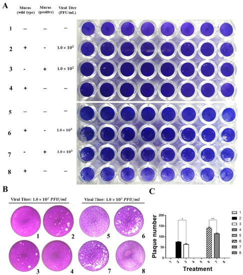
Grass carp IFN1 from mucus inhibited the plaque formation of cultured cells infected with GCRV873. (A,B) Detection of the antiviral ability of grass carp IFN1 from the mucus of transgenic loach. 1, 5: added M199 medium containing 2% FBS; 2: added M199 medium containing 2% FBS and 20% mucus (v/v) from wide type (WT) loaches and infected with GCRV873 (viral titer: 1.0 × 10² PFU/mL); 3: added M199 medium containing 2% FBS and 20% mucus (v/v) from grass carp IFN1-expressing transgenic loaches and infected with GCRV873 (viral titer: 1.0 × 10² PFU/mL); 6: added M199 medium containing 2% FBS and 20% mucus (v/v) from WT loaches and infected with GCRV873 (viral titer: 1.0 × 10² PFU/mL); 7: added M199 medium containing 2% FBS and 20% mucus (v/v) from grass carp IFN1-expressing transgenic loaches and infected with GCRV873 (viral titer: 1.0 × 10³ PFU/mL); 4, 8: added M199 medium containing 2% FBS and 20% mucus (v/v) from WT loaches. (C) Graph of plaque numbers of the Figure 5A. The experiments were performed in triplicate. Data were expressed as mean ± SD. * p < 0.05; ** p < 0.01. |