
Figure 4
- ID
- ZDB-FIG-210128-138
- Publication
- Zhou et al., 2021 - Development of a Mucus Gland Bioreactor in Loach Paramisgurnus dabryanus
- Other Figures
- All Figure Page
- Back to All Figure Page
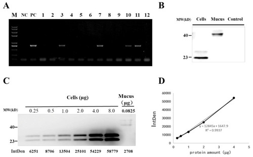
Detection of grass carp IFN1 in transgenic loach. (A) Detection of the IFN1-expressing cassette in F1 offspring by PCR. NC: negative control; PC: positive control; 1–12: F1 loaches. (B) Detection of grass carp IFN1 in mucus using western blots. Cells: the supernatant medium of cultured 293T cells transfected with pcDNA-IFN1-His; Mucus: the mucus collected from F1 loaches containing IFN1-expressing cassette; Control: the mucus collected from wide type loaches. (C) Quantitative analysis of grass carp IFN1 in mucus using western blots. Cells: Purified protein from the supernatant medium of 293T cells transfected with pcDNA-IFN1-His; Mucus: the mucus collected from F1 loaches containing IFN1-expressing cassette. (D) The linear analysis of gray values (IntDen) and protein concentrations in the supernatant medium of 293T cells transfected with pcDNA-IFN1-His to determine the concentration of grass carp IFN1 in the mucus collected from F1 loaches containing IFN1-expressing cassette in (C). |