|
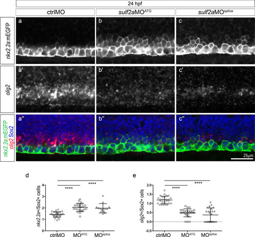
The Nkx2.2a/p3 domain is dorsally expanded at the expense of the Olig2/pMN domain in 24 hpf sulf2a-depleted embryos. Side views of 24 hpf embryos. (a–c”) Double detection of olig2 mRNA and Sox2 in Tg(nkx2.2a:mEGFP) embryos injected with ctrlMO (a–a”), sulf2aMOATG (b–b”) or sulf2aMOsplice (c–c”). Vertical sets present successively the nkx2.2a:mEGFP signal (green in a”–c”), the olig2 mRNA staining (red in a”–c”) and the merged image together with Sox2 staining (blue in a”–c”). Note the dorsal expansion of the nkx2.2a:mEGFP signal associated with a strong decrease in the olig2 signal in sulf2a morphant embryos. (d, e) Quantification of nkx2.2a:mEGFP + /Sox2 + (d) and olig2 + /Sox2 + (e) progenitors in embryos injected with ctrlMO (n = 27), sulf2aMOATG (n = 14) and sulf2aMOsplice (n = 23) from two independent experiments. Datasets were compared with Mann Whitney’s test (two-tailed). Data are presented as a mean number of cells along the dorso-ventral axis per embryo ± s.d (****p < 0.0001).
|