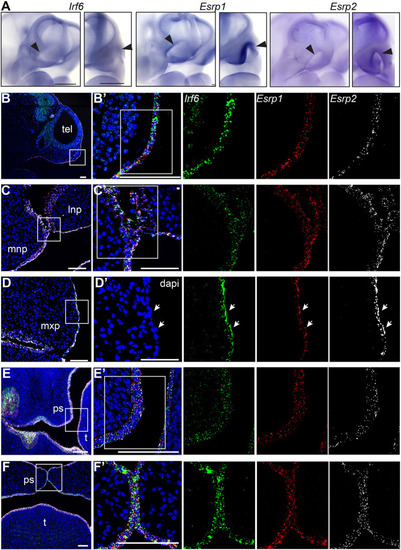
Irf6, Esrp1 and Esrp2 are co-expressed in the oral epithelium of mouse embryos. (A) WISH of E10.5 embryos, showing Irf6, Esrp1 and Esrp2 mRNA expression in the surface epithelium and concentrated within the ectoderm of the frontonasal prominences (arrowheads) and first brachial arch. Oblique and frontal orientation. Scale bars: 500 μm. (B-F′) Sections of E10 (B,B′), E11.5 (C-D′), E13.5 (E,E′) and E15 (F,F′) embryos analyzed by RNAscope ISH, showing mRNA cellular co-expression of Irf6 (green), Esrp1 (red) and Esrp2 (white) in the surface ectoderm (E10), lining the frontonasal and maxillary prominences, including expression in periderm (arrows) (E11.5), and lining the palatal shelves (E13.5, E15). Sagittal (B,B′) and coronal (C-F′) sections; boxed areas are shown at higher magnification in B′, C′, D′, E′ and F′. dapi, 4′,6-diamidino-2-phenylindole; lnp, lateral nasal prominence; mnp, medial nasal prominence; mxp, maxillary prominence; ps, palate shelf; t, tongue; tel, telencephalon. Scale bars: 100 μm.
|
