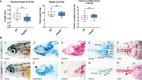
Developmental delay and morphological abnormalities in 20 dpf b3galt6–/– zebrafish. (A) Weight, standard length and Fulton’s condition factor of WT and b3galt6–/– zebrafish larvae at 20 dpf (n = 10). (B) External phenotype of the head of juvenile WT and b3galt6–/– sibling zebrafish. All images are oriented anterior to the left, posterior to the right, dorsal to the top and ventral to the bottom. (1-1’)b3galt6–/– zebrafish display a rounder frontal region (black line), shorter parietal/occipital region (white horizontal line) and a malformed posterior edge of the opercular apparatus (dotted black line), compared to WT control. (2-2’) Representative images of cartilage AB staining of the head. The olfactory region (indicated by “o” in the WT and arrowhead in the b3galt6–/– picture) and the otic region (indicated by white asterisk) show delayed development of cartilage elements in b3galt6–/– zebrafish as compared to WT. (3-3’) Representative images of AR mineral staining of the head. The olfactory region (indicated by “o”) and the otic region (indicated by asterisk) show severely delayed mineralization in b3galt6–/– zebrafish larvae as compared to WT. (4-4’) Representative images of AB staining of the caudal fin. The modified associated elements show developmental delay in b3galt6–/– zebrafish as compared to WT. E, epural; HS PU2, haemal spine of preural 2; HS PU3, haemal spine of preural 3; Hy1–5, hypural 1–5; NS PU2, neural spine preural 2; opc, opistural cartilage; Ph, parhypural (5-5’) Representative images of AR mineral staining of the caudal fin. Caudal vertebrae and their associated elements show no delay in ossification. Malformation or absence of caudal fin associated elements (block arrow indicates a neural arch; arrowhead indicates a missing haemal arch), and malformation of uroneural (uro) and vestigial neural arches and spines (vna/s) are observed in b3galt6–/– zebrafish; hypurals have a bent appearance. Scale bars: images of the head 500 μm, images of the caudal fin 200 μm.
|
